THE LITTLE HEART GUIDE · SEPTEMBER 15, 2026
Different hearts.
A little more understanding.
ASD. VSD. TOF. A handful of letters can feel like a whole new language. Here is a friendly place to start.
What does “congenital” mean?
It means present at birth. A congenital heart defect, or CHD, is a difference in the heart’s structure that can affect blood flow. CHDs range from mild to complex. Nearly 1% of babies born in the United States have a CHD. Source: CDC overview.
SAVE A LITTLE SPACE FOR UNDERSTANDING
Six heart differences,
in plain language.
A selection of common and complex CHDs—not every diagnosis, and not a ranking of severity.
First, the usual route
- Body
- Right heart
- Lungs
- Left heart
- Body
The lungs add oxygen before blood returns to the left side of the heart and travels to the body. The diagrams below zoom in on each difference. Source: NIH.
An opening
Atrial septal defect
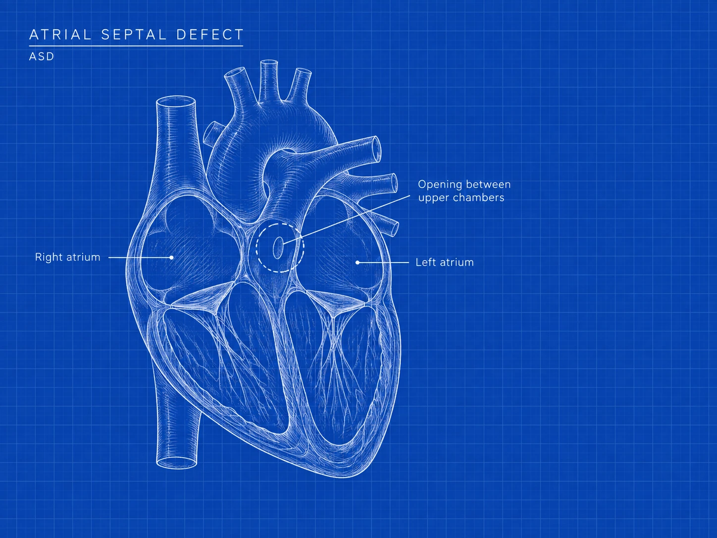
An opening in the wall between the two upper chambers of the heart.
Compare with typical anatomy
An opening
Ventricular septal defect
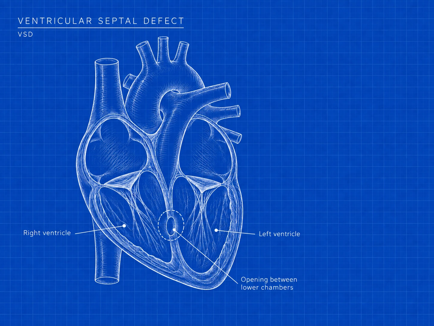
An opening in the wall between the two lower chambers of the heart.
Compare with typical anatomy
A connection stays open
Patent ductus arteriosus
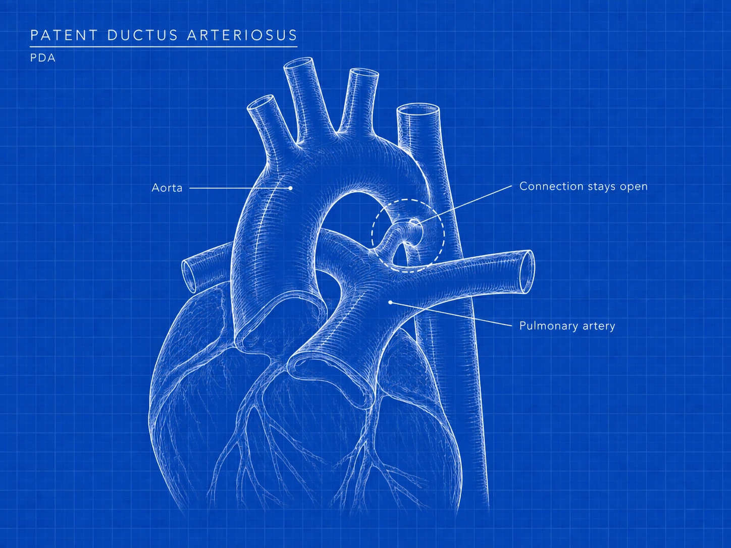
A blood vessel connecting the aorta and pulmonary artery before birth stays open after birth.
Compare with typical anatomy
A narrowed passage
Pulmonary stenosis
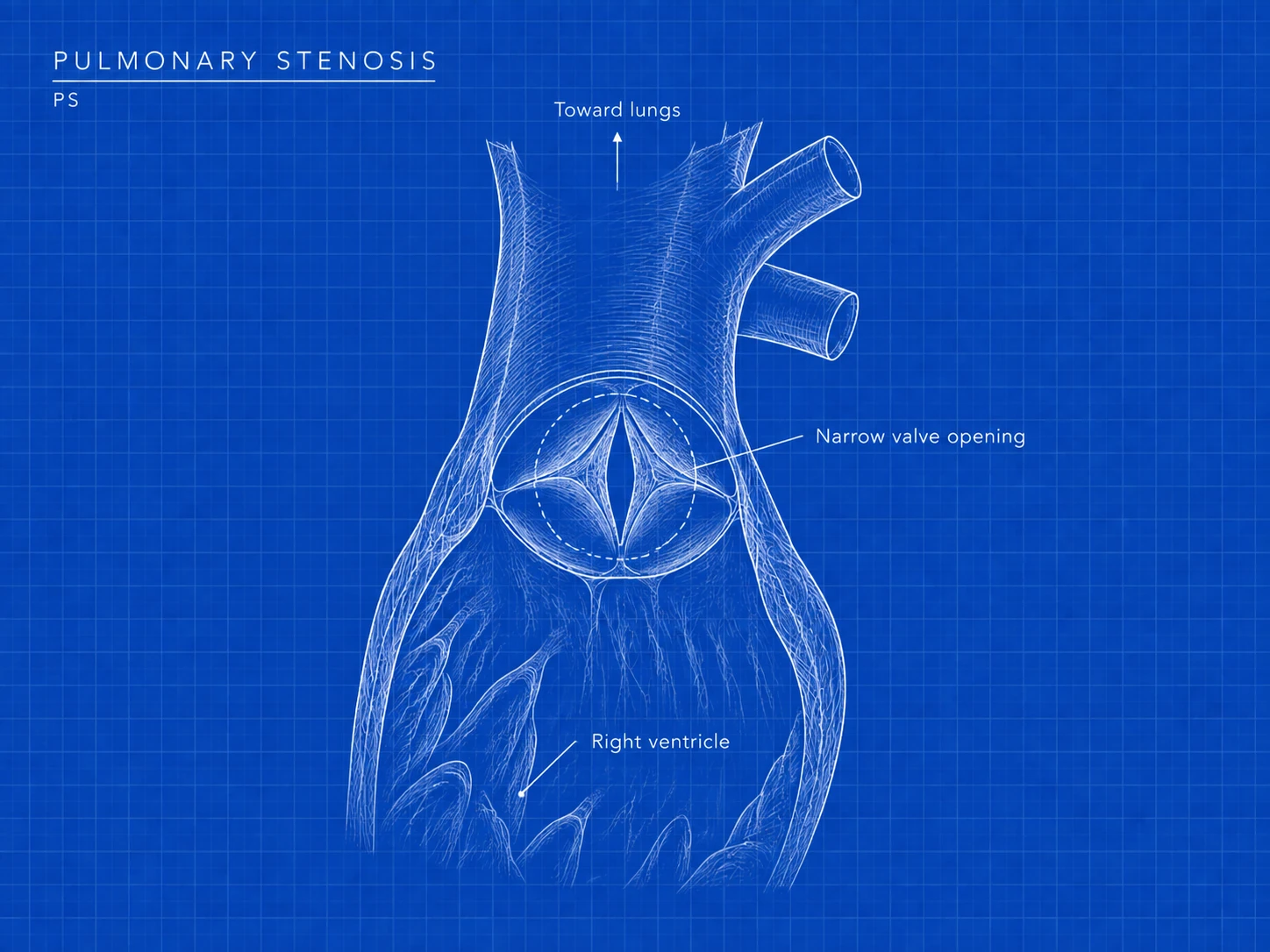
A narrowed or stiff pulmonary valve restricts blood flow from the heart toward the lungs.
Compare with typical anatomy
A narrowed passage
Coarctation of the aorta
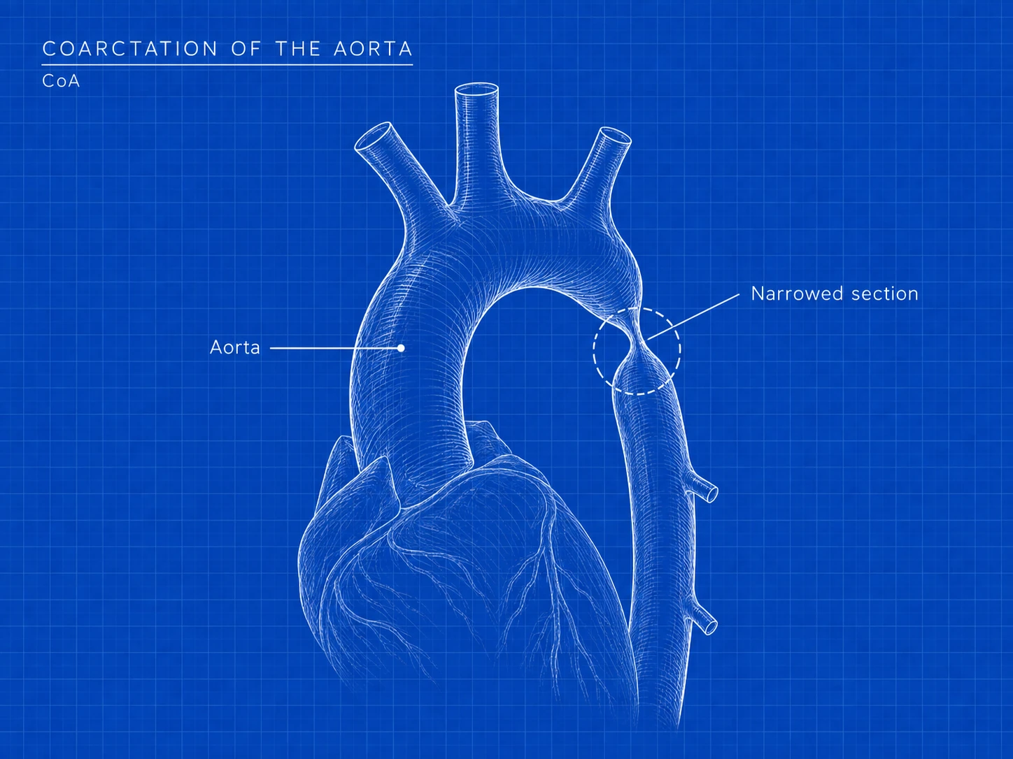
Part of the aorta—the artery carrying oxygen-rich blood to the body—is narrower than usual. The heart must work harder to push blood through.
Compare with typical anatomy
Four differences together
Tetralogy of Fallot
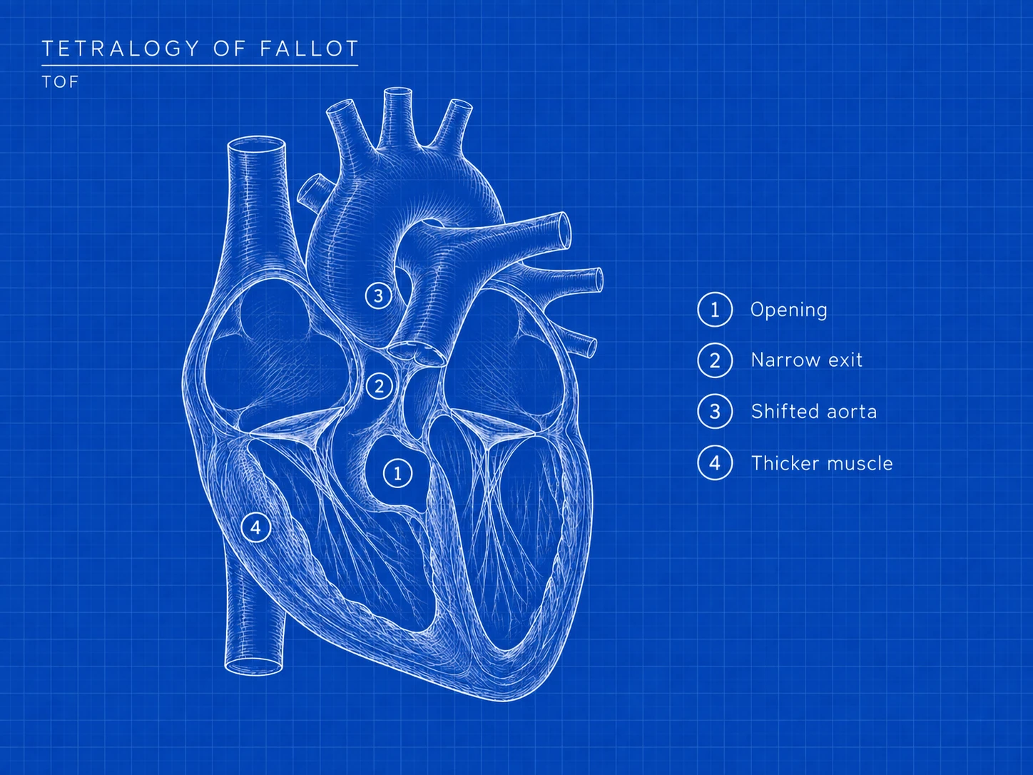
A VSD; narrowing toward the lungs; an aorta positioned over the VSD, receiving blood from both lower chambers; and a thickened right lower chamber muscle. Together, these can reduce the oxygen reaching the body.
- Opening: a VSD between the lower chambers.
- Narrow exit: the route toward the lungs is restricted.
- Shifted aorta: it sits over the VSD and can receive blood from both lower chambers.
- Thicker muscle: the right ventricle’s wall becomes thicker.
Compare with typical anatomy
The same diagnosis can look different from person to person. Your child’s heart team can explain their individual anatomy and care.
A repair is part of a longer story.
Better treatment means more people with CHDs are living longer. But a repair does not always mean a cure; ongoing heart care can still matter. Some defects have few noticeable symptoms, and some are found before birth or through newborn screening. Source: CDC overview.
Three questions to bring along
- Can you draw my child’s heart and show me how the blood flows?
- What follow-up does this particular diagnosis need?
- Which changes should prompt a call to our care team?
You do not have to learn every abbreviation today. Start with the one in front of you, ask the next question, and keep this guide nearby.
Meet the books ↗